This lecture explores advanced methodologies in the field of veterinary anatomical science, focusing on three key areas: novel approaches to bone maceration and skeletal preparations, modern preservation techniques for anatomical specimens, and the integration of digital technologies in anatomical modeling. Dr. Ors Petnehazy will present optimized protocols for efficient and high-quality skeletal preparations, including environmentally conscious and minimally invasive maceration methods. The session will also cover a variety of specimen preservation strategies—ranging from wet specimen conservation to corrosion casting—highlighting their relevance in anatomical education. Finally, the lecture will delve into the digital reconstruction of complex anatomical structures through CT imaging, 3D rendering, and model printing, showcasing workflows from data acquisition to physical modeling. Together, these topics reflect the evolving intersection of traditional anatomy and modern innovation, offering valuable insights for educators, researchers, and veterinary professionals.
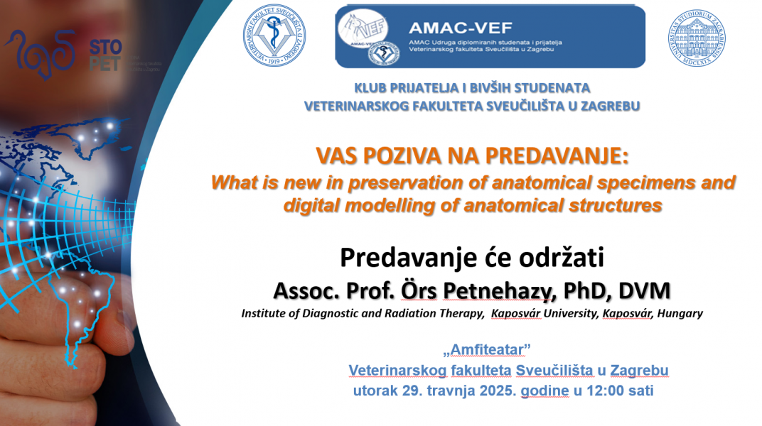
Dr. Ors Petnehazy is an accomplished veterinary anatomist with extensive experience in anatomical education, research, and anatomical specimen development. Currently serving as Associate Professor of Livestock Anatomy at the Hungarian University of Agriculture and Life Sciences, Kaposvár Campus, Dr. Petnehazy has built a diverse academic and professional career spanning multiple institutions and countries.
From 1999 to 2005, he held the position of Practical Teacher and Lecturer at the University of Veterinary Medicine, Budapest. He later joined the University of Alaska Fairbanks as Assistant Professor of Veterinary Anatomy and Diagnostic Imaging (2014–2015), where he contributed to the design of the new veterinary curriculum. From 2015 to 2018, he worked as a Research Veterinarian at the Institute for Diagnostic Imaging and Radiation Oncology, Kaposvár University.
Dr. Petnehazy possesses comprehensive teaching experience, delivering lectures and practical sessions in veterinary anatomy, and is highly dedicated to the maintenance and development of anatomical teaching specimens, including wet preparations, skeletons, and corrosion casts. He is an innovator in anatomical visualization, having produced numerous 3D reconstructions and 3D-printed anatomical models. His CT-based corrosion cast model of a French Bulldog’s head and neck earned recognition at the 2015 “What to Print in 3D” event.
His anatomical specimen photography has been featured in the renowned 5th edition of Dyce, Sack, and Wensing’s Textbook of Veterinary Anatomy.
In addition to his academic work, Dr. Petnehazy is the founder and owner of Justanatomy Ltd., a company specializing in private veterinary anatomical education. Through this platform, he performs cryomacrosectioning, skeletal and vascular preparations, corrosion casting, and the development of anatomical models embedded in clear resin for educational purposes.

 Sveučilište u Zagrebu